BrachyAtlas
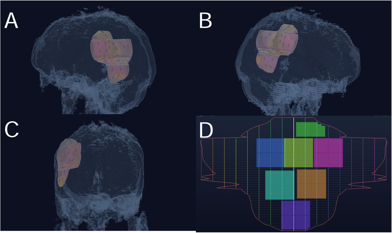
Patient-specific intracranial brachytherapy planning that brings anatomy, imaging, implant geometry and radiation dose into one browser workflow.
I work across clinical AI, computational anatomy, bioelectronic medicine, neuroimaging and experimental neuroscience. My work usually starts with a concrete problem in a clinical or research workflow and ends as a dataset, analysis pipeline, interactive tool, device concept or publication that other people can use.
Open a project directly, or preview the live application without leaving this page.
Patient-specific intracranial brachytherapy planning that brings anatomy, imaging, implant geometry and radiation dose into one browser workflow.
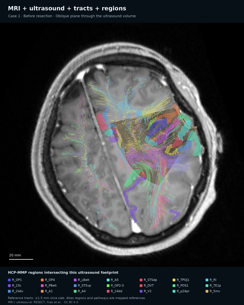
Interactive tools for translating patient-specific connectomic abnormalities into interpretable phenotypes and longitudinal trajectories in brain-tumor research.
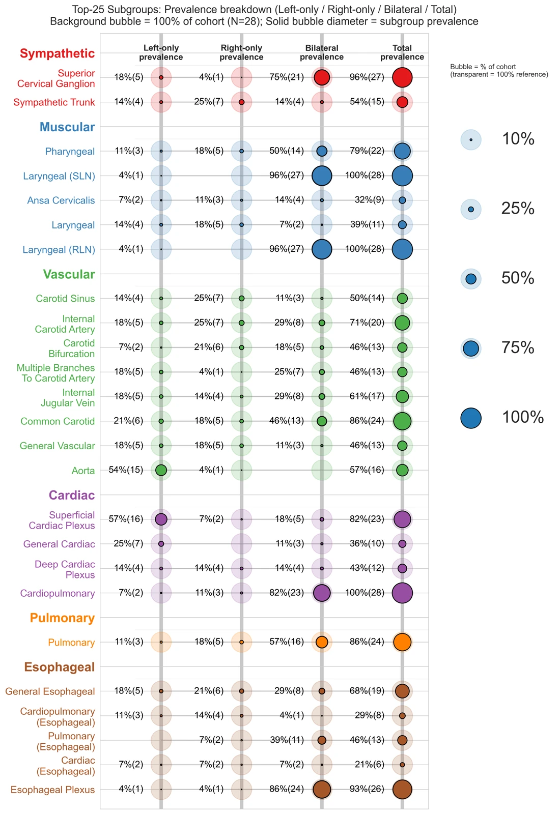
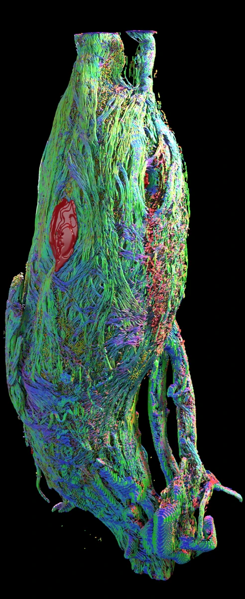
A standardized, surgically relevant map of organ-specific branch emergence from the human vagus nerve, with open data, reproducible analysis and an interactive explorer.
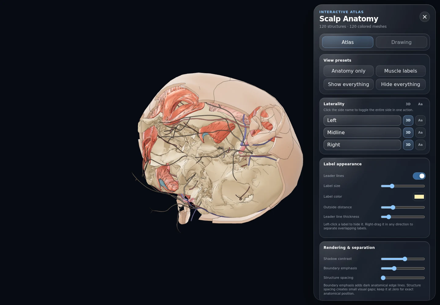
AI-assisted generation of publication-quality anatomy illustrations for scientific and educational use, developed for work with the Society for Neuroplastic Surgery.
A system for organizing long-running AI conversation histories into searchable maps of projects, themes and research development.
Independent work on computational brain mapping, tractography, automated MRI analysis, 3D visualization and cloud-based neuroimaging workflows.
The broader work behind the public tools.
Human cadaveric anatomy, tissue clearing, light-sheet microscopy, single-fiber reconstruction, molecular/spatial profiling and quantitative mapping for selective bioelectronic medicine. Part of NIH-supported human vagus mapping work at the Feinstein Institutes.
Built workflows for intact human nerve and ganglion imaging, including custom holders, mounting systems, access tools and 3D-printed research devices. Some device concepts became the basis for provisional patent work.
Computational methods for cranial reconstruction, including automated 3D deformity measurement, transcranioplasty ultrasound through sonolucent implants, and anatomy/education tools for the Society for Neuroplastic Surgery.
Clinical evaluation of language models, AI-assisted research workflows, and an invited review for Neurosurgery on data quality, leakage, validation, interpretability, reproducibility and clinical relevance in neurosurgical AI studies.
Work spanning normal-pressure hydrocephalus, connectomics, tractography, cranial morphology, AR/head-mounted fiber tractography, 7T ex-vivo imaging and imaging-to-operative-planning workflows.
Elmezzi Scholar and PhD candidate in Bioelectronic Medicine. Human vagus and nodose mapping, peripheral-nerve imaging, computational anatomy and translational device work.
Attending neurosurgeon after residency, managing emergency neurosurgical and trauma care in a public hospital.
Comprehensive neurosurgical training with 450+ operations. Built brain-mapping, tractography and quantitative imaging workflows alongside clinical practice.
Frontline emergency care, triage, resuscitation, long-distance transfers and regional EMS coordination in Muş, Türkiye.
Python, LLM application development, model evaluation, evaluation harnesses, tool use, agentic workflows, RAG/embeddings, computer vision, deep learning, SQL, APIs, deployment and interactive visualization.
MRI, CT, DTI/fMRI, tractography, connectomics, computational anatomy, 3D reconstruction, radiomics, quantitative morphology and operative planning.
Light-sheet microscopy, tissue clearing, immunohistochemistry, molecular/spatial anatomy, human cadaveric tissue processing, segmentation and fiber tracking.
3D printing, CAD-style modeling, surgical guides, anatomical access modeling, prototype iteration, cadaveric testing and surgeon-engineer workflow translation.